
A new milestone for science at NTU: The inauguration of the Max Planck-IAS-NTU Center
瀏覽器版本過舊,或未開啟 javascript
請更新瀏覽器或啟用 javascript
Spotlights
Date: Dec 28, 2018
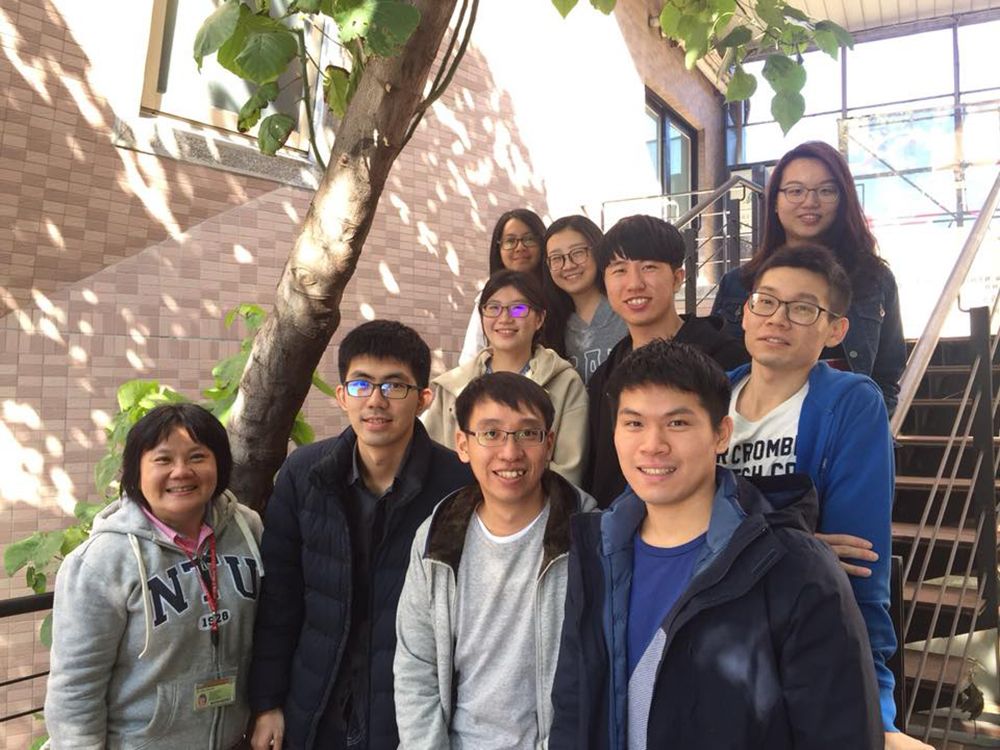
Lab members led by Associate Prof. Chun-Liang Pan.
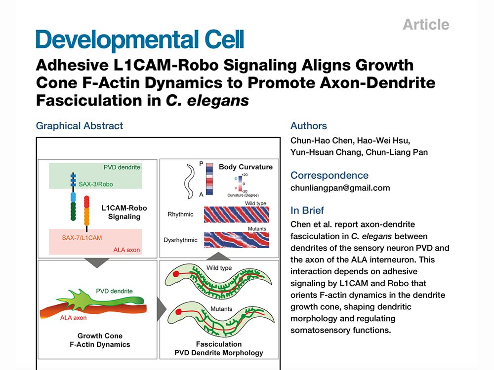
Associate Prof. Chun-Liang Pan’s team published its latest study in the prestigious journal, Developmental Cell.
How does a neuron build its sophisticated architecture that enables the many intricate functions of the nervous system? A new study published by the lab of Chun-Liang Pan (潘俊良), Associate Professor at the NTU Institute of Molecular Medicine, finds that bundling between dendrites and axons, two distinct neuronal processes that have unique molecular compositions, can sculpt the fine morphology of dendritic arborization. This dendrite-axon bundling is important for animals to properly sense mechanical stimuli from both the exterior and interior environment. This is the first evidence that dendrites form fascicles, or bundles, with axons. Pan and Chun-Hao Chen (陳俊豪), postdoc and first author of the paper, reported their findings in the journal, Developmental Cell.
Dendrites are neuronal processes that receive information to enable neural computation. The numerous dendrites of a neuron do not grow in a random fashion. Rather, they mostly follow certain rules such that more peripheral branches come successively from proximal dendrites. While the gross morphology of dendritic arborization has been thought to be largely determined by genetic programs that specify neuronal types, evidence emerges that interactions between neurons also shape dendrite morphology. Different axons of the same neuronal type can bundle together to facilitate their projection to other brain areas, but it is not known whether dendrites also bundle with axons to foster their development.
The Pan lab addressed this question using Caenorhabditis elegans, a tiny roundworm (nematode) that has become a model organism for neurobiology. In C. elegans, the PVD neuron detects mechanical stimuli applied on the worm’s skin surface and presumably also the muscle tension generated during its crawling. The proximal dendrites of PVD bundle with the axon from another neuron, ALA. Chen found that PVD dendrites went wrong when ALA or its axon was disrupted using genetic tricks. By systemically investigating molecules that may have adhesive functions and are predicted to be on the membrane of PVD and ALA, Chen identified SAX-7/L1CAM and SAX-3/Robo as a ligand-receptor pair that bundled ALA axons and PVD dendrites. SAX-7 on the ALA membrane interacts with SAX-3 on PVD dendrites; in the absence of either SAX-7 or SAX-3, PVD dendrites wandered about and resulted in aberrant dendritic arborization.
Using high-speed fluorescence microscopy, Chen produced real-time images that documented the dynamics of F-actin, a major protein that supports cell growth and movements, when PVD dendrites grew actively. The images suggested that adhesion between ALA and PVD helped to align F-actin dynamics with the directional extension of the dendrites. Interestingly, when ALA-PVD bundling was disrupted, the worm had a problem detecting mechanical stimulation, and it also failed to generate highly rhythmic waveforms during locomotion, two functions that the PVD neuron serves. These findings indicate that bundling with an axon is crucial for the dendrites to have proper morphology and functions.
Chen obtained his PhD degree under the mentorship of Pan, and conducted this research as a postdoc in the lab. Supported by a postdoctoral fellowship from the Ministry of Science and Technology (MOST), Chen is now at the California Institute of Technology to continue his journey into neuroscience. Pan attributes the success of this project to Chen's talents and passion for science, as well as the imaging expertise of Hwa-Man Hsu (徐華蔓) at the First Core Lab of the NTU College of Medicine. Other authors include Hao-Wei Hsu (許皓瑋) and Yun-Hsuan Chang (張運玄), both graduate students of the NTU Institute of Molecular Medicine.
Reference:
Chun-Hao Chen, Hao-Wei Hsu, Yun-Hsuan Chang, and Chun-Liang Pan. “Adhesive L1CAM-Robo Signaling Aligns Growth Done F-Actin Dynamics to Promote Axon-Dendrite Fasciculation in C. elegans.” Developmental Cell, Dec 13, 2018. DOI: https://doi.org/10.1016/j.devcel.2018.10.028.
(Source: Associate Prof. Chun-Liang Pan, NTU Institute of Molecular Medicine)
A new milestone for science at NTU: The inauguration of the Max Planck-IAS-NTU Center
A Distinguished Global Research Center Established at NTU under Trilateral Cooperation
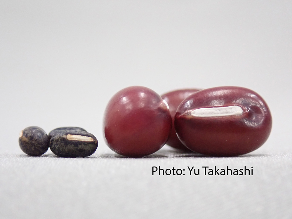
Collaborative study between NTU and Japan uncovers the origin of Adzuki Beans and agriculture in Japan
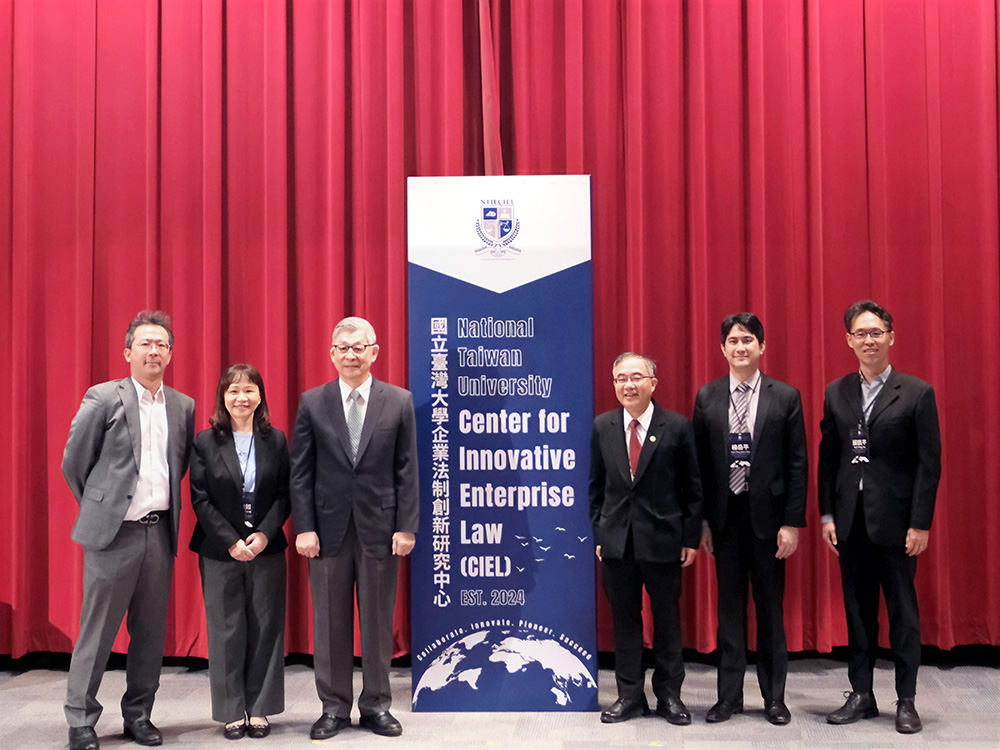
NTU Launches Center for Innovation in Enterprise Law—with Forum Highlighting Trump’s Policy and Legal Shifts Amid Geopolitical Tensions

NTU and Ministry of Environment Sign MOU to Advance Net-Zero Transition and Environmental Resilience
Current Spotlights
A new milestone for science at NTU: The inauguration of the Max Planck-IAS-NTU Center
A Distinguished Global Research Center Established at NTU under Trilateral Cooperation
Collaborative study between NTU and Japan uncovers the origin of Adzuki Beans and agriculture in Japan
NTU Launches Center for Innovation in Enterprise Law—with Forum Highlighting Trump’s Policy and Legal Shifts Amid Geopolitical Tensions
NTU and Ministry of Environment Sign MOU to Advance Net-Zero Transition and Environmental Resilience