


A new milestone for science at NTU: The inauguration of the Max Planck-IAS-NTU Center
瀏覽器版本過舊,或未開啟 javascript
請更新瀏覽器或啟用 javascript
Spotlights
Date: Apr 24, 2015
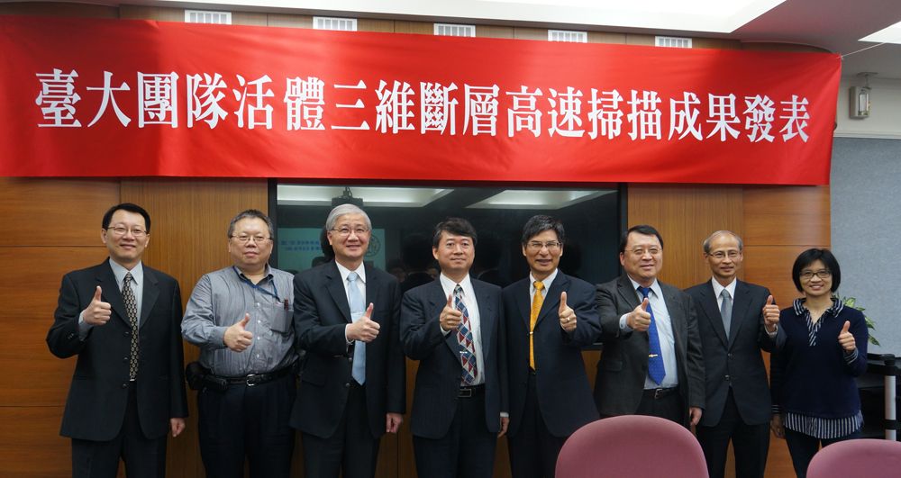
NTU announces licensing of the in vivo high speed and submicron 3D OCT technique to Apollo Medical Optics.
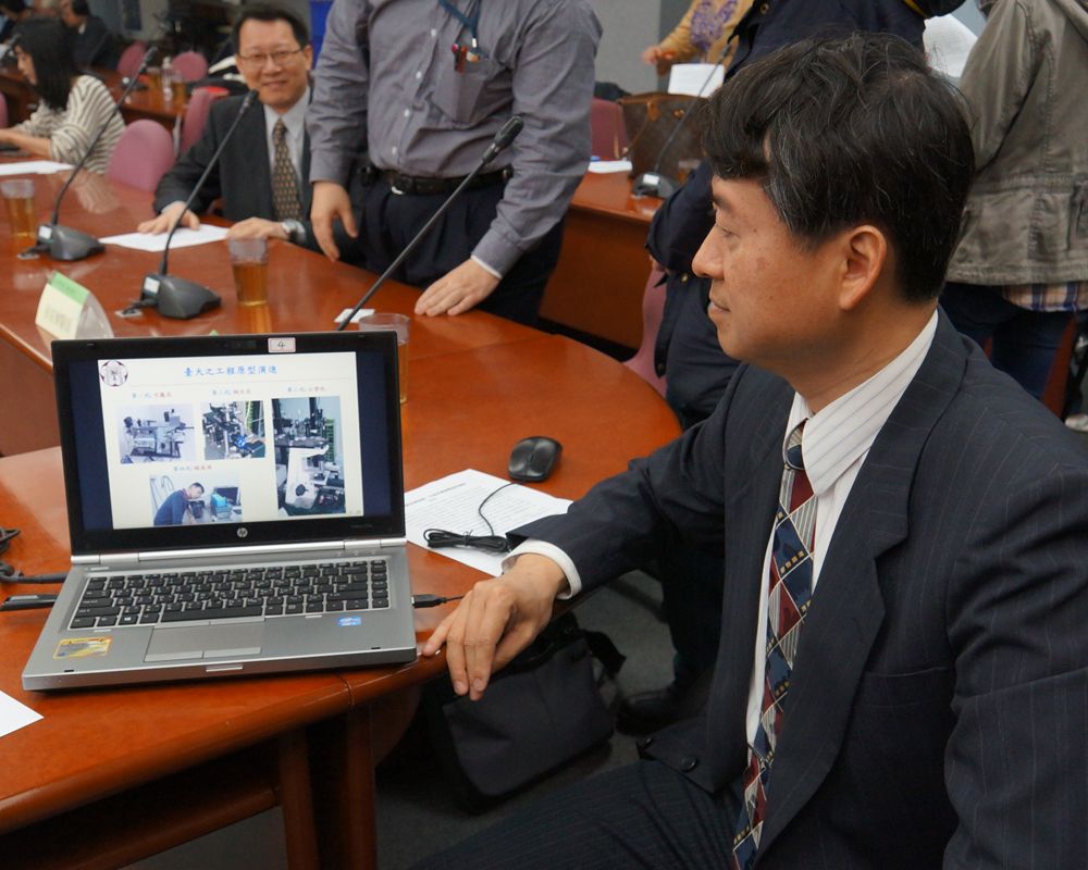
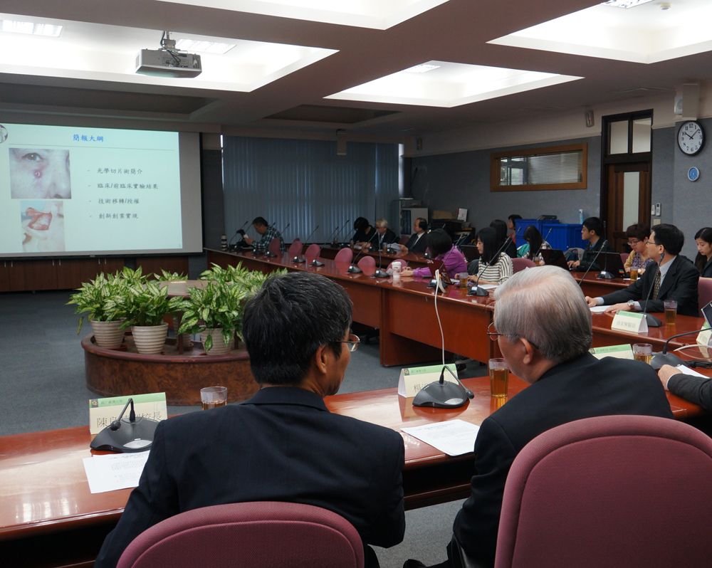
Prof. Sheng-Lung Huang explains the project to the press.
The technique is expected to reduce diagnosis time and medical costs.
Imagine a world where the time, money, and efforts spent on detecting such diseases as cancer is significantly cut. This may well become a reality in the near future with the development of a non-invasive in vivo technique developed by a team from National Taiwan University.
In Taiwan as well as in other developed countries, health care expenditure takes up as much as 15-20 percent of the national GDP, and medical costs continue to escalate with increasingly aging societies. The in vivo and high speed 3D tomography medical project developed by Prof. Sheng-Lung Huang (黃升龍) of the Department of Electrical Engineering may be the answer to this concern as it is capable of reducing the time and costs needed for diagnosis of diseases and cancers.
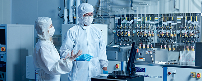
Equipped with cellular resolution, the in vivo high speed and submicron 3D optical coherence tomography (OCT) technique can perform quantitative analysis of a single cell with measurements of just 0.01 mm. This is at least 100 times smaller than what present devices can detect. Using light sources to penetrate the skin, tissue anatomy and the flow of blood cells in micro-vessel can also be imaged in real time. This “optical biopsy” method may replace the conventional time-consuming practice of conducting physical biopsies. These are the major factors that allow for early stage diagnoses to take place. The core technology behind the NTU project is the inclusion of proprietary high-brightness crystalline fibers. The fibers can produce broadband emissions that range from visible light to near infrared in achieving high speed and high resolution anatomical imaging and dynamic blood flow analysis. At present, the technology has entered into clinical and preclinical trials, as well as initial phases of animal testing for research on skin cancer, colon cancer, and age-related macular degeneration.
Initial findings from the trials have shown that the project’s optical biopsy technique, which features sub-micron resolutions in both the lateral and axial directions, are extremely likely to replace physical biopsies for early diagnosis of diseases and cancers in the near future. The project has been more than once featured in the website of the internationally acclaimed Optical Coherence Tomography News.
Prof. Huang points out that the project has entered into its tenth year of development with support from the Ministry of Science and Technology and the Ministry of Economic Affairs. The release of its fourth-generation prototype has a total of 15 awarded and pending USA patents. To further expedite the research, the project’s intellectual property rights have been licensed to Apollo Medical Optics for product development and medical certification. The technique is expected to soon be used in domestic hospitals in the departments of dermatology, pediatrics, immunology, and rheumatology.
A new milestone for science at NTU: The inauguration of the Max Planck-IAS-NTU Center
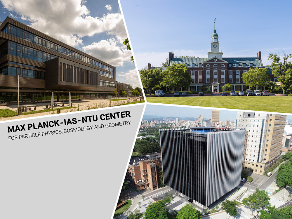
A Distinguished Global Research Center Established at NTU under Trilateral Cooperation
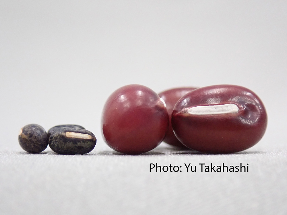
Collaborative study between NTU and Japan uncovers the origin of Adzuki Beans and agriculture in Japan
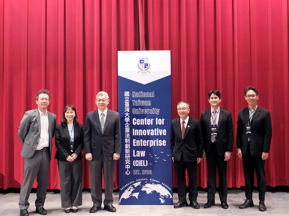
NTU Launches Center for Innovation in Enterprise Law—with Forum Highlighting Trump’s Policy and Legal Shifts Amid Geopolitical Tensions
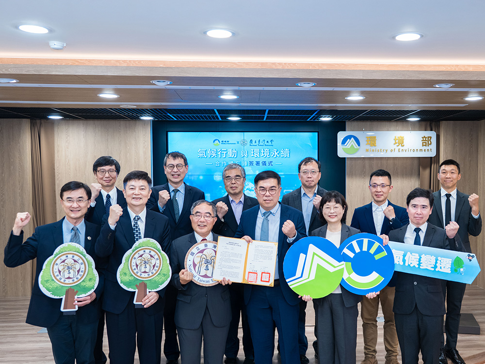
NTU and Ministry of Environment Sign MOU to Advance Net-Zero Transition and Environmental Resilience
Current Spotlights
A new milestone for science at NTU: The inauguration of the Max Planck-IAS-NTU Center
A Distinguished Global Research Center Established at NTU under Trilateral Cooperation
Collaborative study between NTU and Japan uncovers the origin of Adzuki Beans and agriculture in Japan
NTU Launches Center for Innovation in Enterprise Law—with Forum Highlighting Trump’s Policy and Legal Shifts Amid Geopolitical Tensions
NTU and Ministry of Environment Sign MOU to Advance Net-Zero Transition and Environmental Resilience