


A new milestone for science at NTU: The inauguration of the Max Planck-IAS-NTU Center
瀏覽器版本過舊,或未開啟 javascript
請更新瀏覽器或啟用 javascript
Spotlights
Date: Apr 8, 2021
Cell-Free Virus-Host Chimera DNA From Hepatitis B Virus Integration Sites as a Circulating Biomarker of Hepatocellular Cancer.
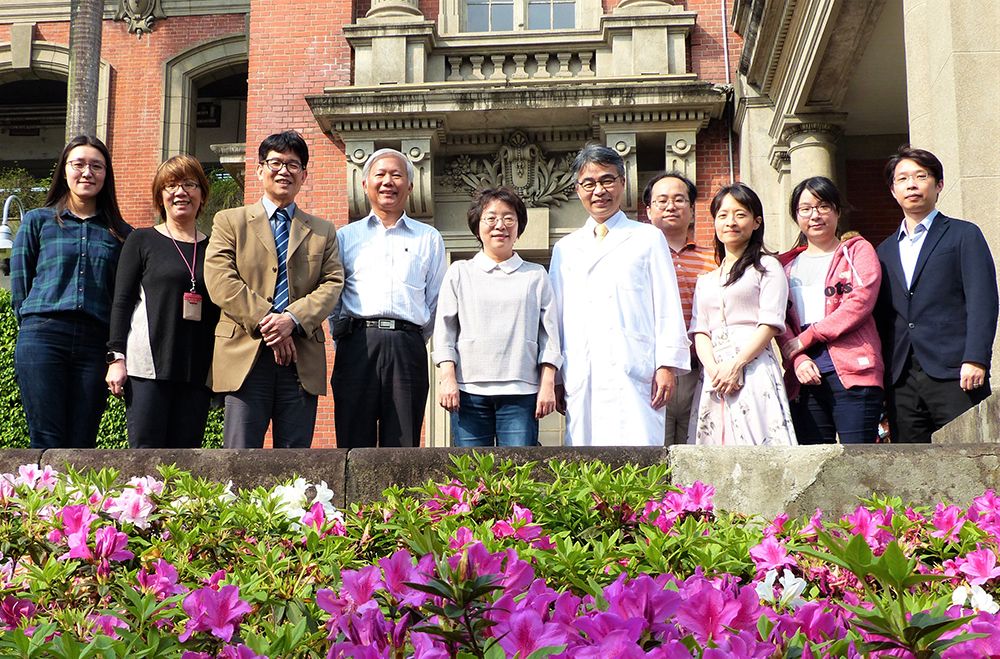
NTU Hepatitis Research Team.
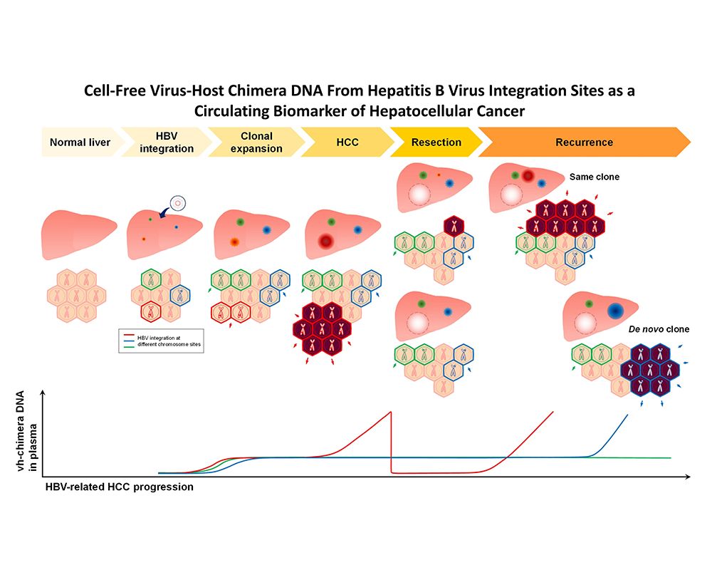
When hepatocytes are infected with hepatitis B virus (HBV), about 0.1% of them take HBV DNA into their chromosome at random sites. Unique virus-host chimera DNAs (vh-DNA) are generated from junctions of random HBV integrations in the genome. The clonal expansion of hepatocytes on their path to malignancy causes most tumor cells to have their own unique vh-DNA, which becomes a specific signature tumor marker for the individual HCC in question. As vh-DNA is present in about 90% of HBV-related HCC cases and vh-DNA is released in circulation during tumor turnover, detection of vh-DNA from the tumor in plasma is potentially a novel blood tumor marker for HBV-related HCC.
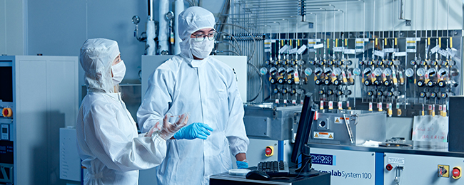
The hepatitis research team at NTU College of Medicine, led by Academia Sinica Academician Pei-Jer Chen (陳培哲), including Professor Shiou-Hwei Yeh (葉秀慧) of the Department of Microbiology, Professor Ming-Chih Ho (何明志) of the Department of Surgery at NTU Hospital and researchers at TCM Biotech International Corp. (泰宗生物科技公司), has successfully validated the potential of the virus-host chimera DNA (vh-DNA) as a circulating biomarker for HCC. Fifty patients with resectable HBV-related HCC were recruited for the study. HBV DNA integration was detected in the resected tumor tissues in about 90% of the subjects using capture-NGS platform technology. A vh-DNA-specific droplet digital PCR (ddPCR) assay was designed based on the detected HBV integration sequence for individual HCC, which showed a positive correlation between the copy numbers of vh-DNA in plasma samples and the tumor sizes. The detection limit of vh-DNA for HCC is approximately 1.5 cm in diameter. The positive detection rate was significantly higher than that of AFP and PIVKA-II, which are the serum tumor markers most widely used in the current clinical setting. The results support the conclusion that vh-DNA can be used as a potential blood tumor marker for the detection of HBV-related HCC.
Since about one-third of HCC patients develop recurrent tumor in the first year after tumor resection, in association with poor survival rates, the research team further attempted to use this technology to detect vh-DNA in the blood samples collected from patients two months after surgery to verify whether it could be used as a biomarker for tumor recurrence. Cell-free DNA in the blood has low stability with a half-life of only a few hours. Therefore, vh-DNA detected in the blood collected two months after tumor resection would indicate that tumor cells are still present in the patient’s body and suggest a high probability for tumor recurrence. The results of the study show that 90% of the patients with positive vh-DNA in the blood two months after tumor resection experienced HCC recurrence within one year. The statistical analysis clearly indicates that the “presence of vh-DNA in plasma collected two months after tumor resection” is the most significant risk factor for HCC recurrence within one year. This study supports the feasibility of applying vh-DNA clinically to detect the postoperative recurrence of HBV-related HCC. All that required is collecting blood samples for recurrence monitoring, which will help physicians provide appropriate treatment to patients as early as possible. This part of the study has currently entered the clinical trial stage with collaboration from the industry.
In addition to providing a novel and clinically practical blood tumor marker for detecting HBV-related HCC, the identification of unique HBV integration sites in the HCC genome can also clarify the clonality of the recurrent tumor relative to the primary tumor as well as provide clues for further tumor treatment. The results of this study have been published in the journal Hepatology, and the paper has also been recommended by F1000Prime (the editorial team of the F1000 Gastroenterology & Hepatology Faculty) for its “special significance in its field”, which is an important recognition of the study’s contribution.
Refer to the following for the study’s full text: https://aasldpubs.onlinelibrary.wiley.com/doi/full/10.1002/hep.31230
A new milestone for science at NTU: The inauguration of the Max Planck-IAS-NTU Center
A Distinguished Global Research Center Established at NTU under Trilateral Cooperation
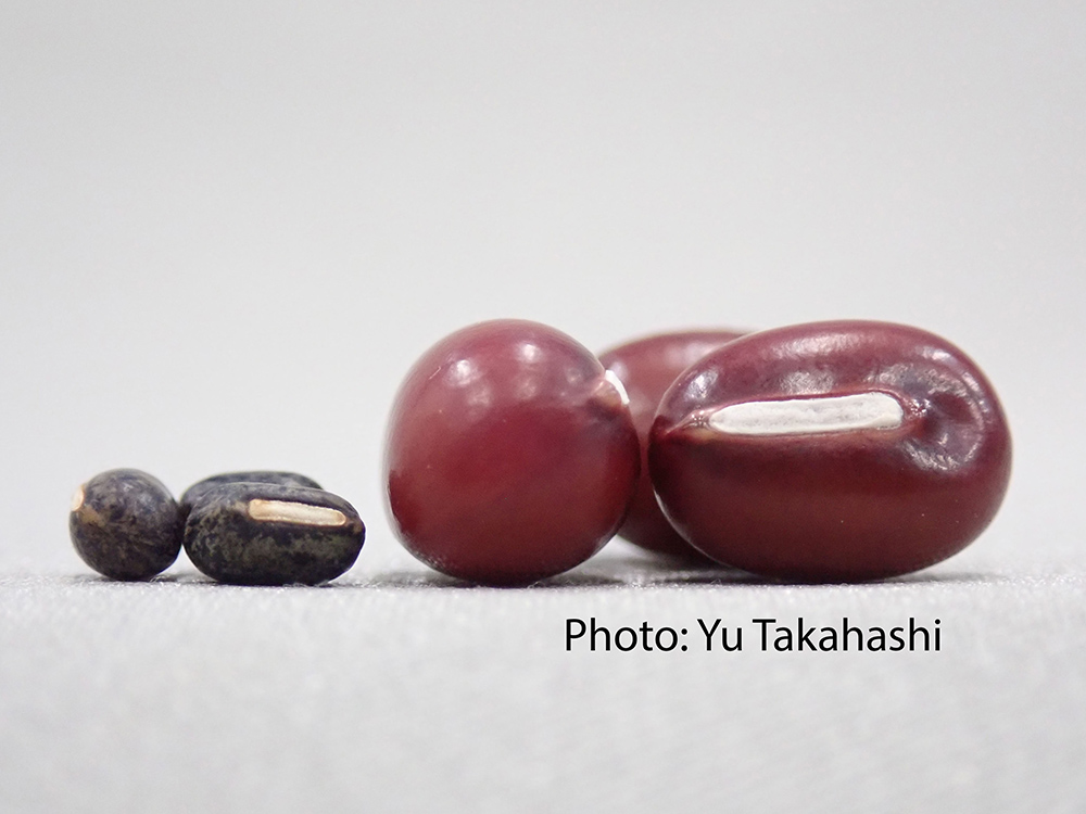
Collaborative study between NTU and Japan uncovers the origin of Adzuki Beans and agriculture in Japan
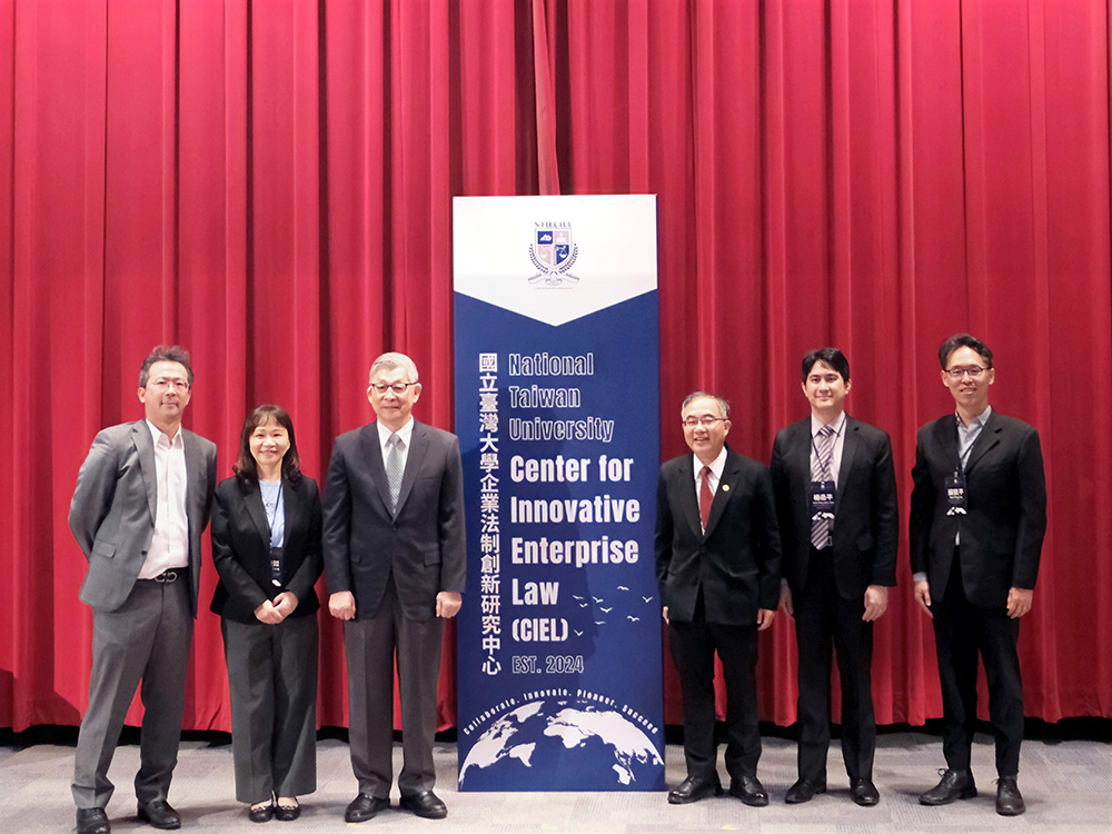
NTU Launches Center for Innovation in Enterprise Law—with Forum Highlighting Trump’s Policy and Legal Shifts Amid Geopolitical Tensions

NTU and Ministry of Environment Sign MOU to Advance Net-Zero Transition and Environmental Resilience
Current Spotlights
A new milestone for science at NTU: The inauguration of the Max Planck-IAS-NTU Center
A Distinguished Global Research Center Established at NTU under Trilateral Cooperation
Collaborative study between NTU and Japan uncovers the origin of Adzuki Beans and agriculture in Japan
NTU Launches Center for Innovation in Enterprise Law—with Forum Highlighting Trump’s Policy and Legal Shifts Amid Geopolitical Tensions
NTU and Ministry of Environment Sign MOU to Advance Net-Zero Transition and Environmental Resilience